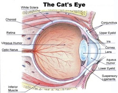
Cat’s have a visual field of view of about 200°. Their eyes work extremely well in low light and, in darkness, cats eyes are able to function in approximately one-sixth of the light needed for human vision. The pupil of a cat’s eye must open up as wide as possible to see in low level lightCat’s also use circular ciliary muscle, but theirs are controlled by two shutter-like ciliary muscles. A characteristic slit-like pupil in bright light conditions is also controlled by this muscle. All cats pupils are therefore elliptical (oval-shaped), but some “big cats” pupils are more circular when dilated (widen).
Cat’s have bigger eyes than human’s. Because of this, more light is able to enter the eye. The lens of cat’s eyes are curved which enables objects to become sharp even at the edges of the cat’s lens. The size of the anterior chamber and the curvature of the cornea is also greater, which helps more light to be refracted (change direction when at an angle) onto the light-sensitive retina.
The tapetum lucidum also enables the amount of light that hits the retina to be increased. This is located behind the retina and it acts as a mirror reflecting light back onto the cat’s sensory cells in the retina. This gives the “glow-like” tent to cat eyes when they are in the dark and a ray of light is caught with them.
There are two distinct types of light receptor cell on the retina. One of which are called Cones, are sensitive to high levels of light. Cones are also used in color vision while Rods are sensitive in low light conditions. In cats, there is a greater concentration of Rods, aiding their night-time vision. In cats, the concentration of receptor cells at the centre of the eye is along a broader, horizontal band. This gives the cat far more sensitivity to movement along the horizontal axis and they are more able to detect prey movement along the ground at greater distances.
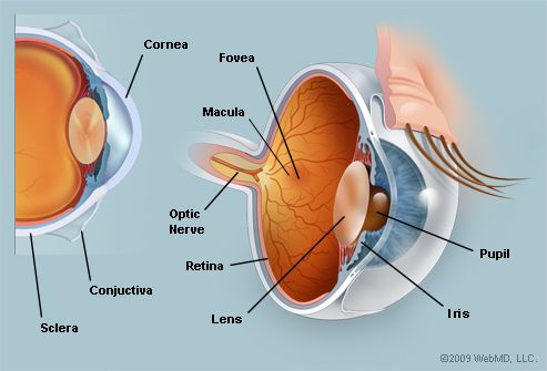
The difference between humans and cats is that humans only have a visual view of 180°. Our eyes work extremely well in bright light and we have a limit to how the size can change since it is controlled by the circular ciliary muscle. In humans, there is a greater concentration of receptor cells at the centre of the eye, leading to the optic nerve. The human eye is a slightly asymmetrical globe and is about an inch in diameter.
(=^● ⋏ ●^=)
